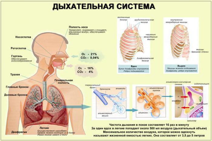
The respiratory system is represented by variousbodies, each of which performs a specific function. In it allocate airways and respiratory part. The latter includes the lungs; the respiratory tract includes the larynx, the trachea, the bronchi and the nasal cavity. The inner part is lined with a cartilaginous skeleton, which is why the tubes do not fall down. Also on the walls there is the ciliated epithelium, cilia, which hold dust and various foreign particles, removing them from the nasal passage along with mucus. Each section of the respiratory system has its own characteristics and performs a specific function.

Nasal cavity
Airways begin with the nasal cavity.This body performs several functions at once: it retains foreign particles that enter the respiratory system together with the air, allows you to hear odors, moisturizes and warms the air.
The nasal cavity is divided into two parts nasalseptum. Choans are located behind, connecting the airway with the nasopharynx. The walls of the nasal passage are formed by bone tissue, cartilage and lined by mucous membrane. Under the influence of irritants, it swells, inflames.
In the nasal passage the largest is the cartilagepartitions. There are also medial, lateral, upper and lower partitions. On the lateral side are three turbinates, between which there are three nasal passages. The upper nasal passage contains a large number of olfactory receptors. The middle and lower parts are considered respiratory.
The initial airways are connected with the paranasal sinuses: maxillary, frontal, ethmoid and sphenoid.

Nasal breathing
During breathing air enters the nose, whereit is cleansing, moisturizing and warming. Then he goes to the nasopharynx and then to the pharynx, where the opening of the larynx opens. In the pharynx, the digestive and respiratory tracts intersect. This feature allows a person to breathe through the mouth. However, in this case, the air, passing through the organs of the airways, is not cleaned.
The structure of the larynx
На уровне шестого и седьмого шейных позвонков the larynx begins. In some people, its visually noticeable slight elevation. During a conversation, coughing of the larynx shifts, following the hyoid bone. In childhood, the larynx is located at the level of the third cervical spine. Older people descend to the level of the seventh vertebra.
Below the larynx enters the trachea. In front of it are the neck muscles, on the sides - the vessels and nerves.
The larynx has a skeleton represented by cartilage.cloth. The cricoid cartilage is located in the lower part, the anterolateral walls are represented by the thyroid cartilage, and the upper opening is covered by the epiglottis. The back of the organ has paired cartilage. In comparison with the front and side, they have a softer structure, due to which they easily change their position relative to the muscles. Behind are the carob, wedge-shaped and scaly cartilages.
In their structure, the airways are similar to many hollow organs: from the inside they are lined with mucous tissue.
The larynx has three divisions:lower, middle and upper. Anatomic complex structure differs middle section. On its side walls is a pair of folds, between which there are ventricles. The lower folds are called vocal folds. In their thickness are the vocal cords, which are formed by elastic fibers and muscles. Between the right and left folds there is a gap, which is called the voice. In men, it is slightly larger than in women.

Trachea structure
A continuation of the larynx is the trachea. This airway is also lined with mucous tissue. The length of the trachea averages ten centimeters. In diameter, it can reach two centimeters.
Стенки органа имеют несколько хрящевых неполных rings that are closed with bundles. The wall behind the trachea is membranous and contains muscle cells. The mucous membrane is represented by the ciliated epithelium and has many glands.
The trachea begins at the level of the sixth cervical vertebra, ends at the level of the fourth or fifth. Here is the division of the trachea into two bronchus. The place of bifurcation is called bifurcation.
Anterior to the trachea is the thyroid gland. Its isthmus is located at the level of the third ring of the trachea. Located behind the esophagus. On both sides of the body are the carotid arteries.
In children, the trachea is blocked in front by the thymus gland.

Structure of the bronchi
From the place of bifurcation of the trachea, the bronchi begin. They depart almost at a right angle and go to the lungs. On the right side the bronchus is wider than on the left.
The walls of the main bronchi have incomplete cartilaginousrings. The organs themselves are divided into medium, small and bronchi of the first, second, third and fourth order. In the small caliber there is no fibrocartilage tissue, and in the middle there is an elastic cartilage tissue, which replaces the hyaline cartilage.
The bronchi of the first order have a branch in the lungs to the lobar bronchi. They are divided into segmental and further lobular. From the last acini depart.
Lung structure
The airway completes the lungs, whichare the largest organs of the respiratory system. They are located in the chest. On both sides of them are the heart and large vessels. Around the lungs is the serous membrane.

The lungs have a cone shape with a base directed toward the diaphragm. The top of the body is located three centimeters above the clavicular bone.
In the human lung there are several surfaces: the base (diaphragmatic), costal and medial (mediastinal).
Bronchi, blood and lymph vessels enterinto the lungs through the mediastinal surface of the organ. They form the root of the lung. Next, the body is divided into two lobes: left and right. At the anterior margin of the left lung there is a heart fossa.
Lobes of each lung consist of smallsegments, among which is bronchopulmonary. Segments have the form of pyramids, the base of which is facing the surface of the lung. Each body has ten segments.
Bronchial tree
The lung area, which is somewhat separated fromneighboring special layer, called the bronchopulmonary segment. The bronchi of this area strongly branched. Small elements with a diameter of no more than a millimeter enter the lung lobe, and branching continues inside. These small parts are called bronchioles. They are of two types: respiratory and terminal. The latter are characterized by a transition to the alveolar passages, and those are completed with alveoli.
The whole complex of branching bronchus is called a bronchial tree. The main function of the airways is gas exchange between the air and blood filling the alveoli.
Pleura
The pleura is a serous membrane.lung. It covers the organ from all sides. The shell runs along the edge of the lungs to the chest, forming bags. Each lung has its own individual shell.
There are several types of pleura:
- Pristyonochnaya (it is lined with the walls of the chest cavity).
- The diaphragmatic.
- Mediastinal.
- Ribbed
- Pulmonary.
Between the pulmonary and parietal pleura is the pleural cavity. It contains fluid that helps reduce friction between the lungs and the pleura during respiratory movements.
Легкие и плевра имеют разные границы.At the pleura, the upper border is three centimeters higher than the first rib, and the rear border is at the level of the twelfth rib. The anterior border is variable and corresponds to the line of transition of the costal pleura to the mediastinal.
Pneumatic organs perform the respiratory function. Without the organs of the respiratory system is impossible to live.