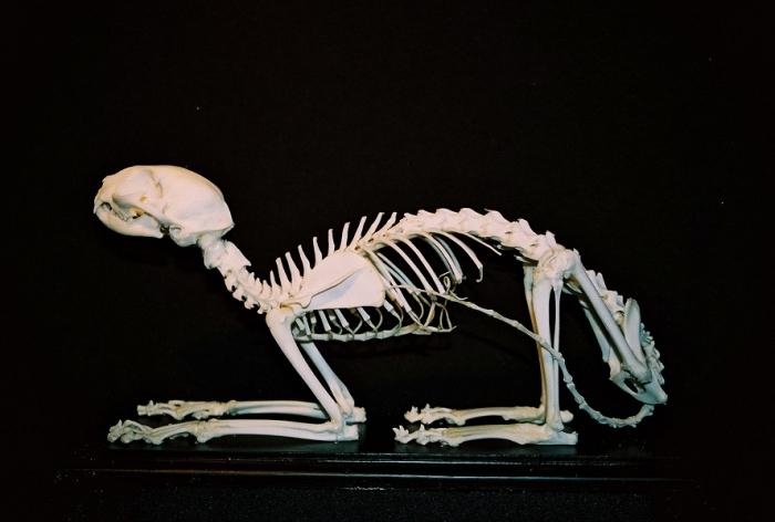
Everybody can imagine a skeletonman, thanks to the numerous photographs and drawings that each of us saw in school. But do we know that the skeleton of an adult person consists of a large number of different bones, each of which performs a specific function?
The human skeleton: what is it made of?
The skeleton of a person is his support. It is not only able to act for the human body as a repository for its internal organs and systems, but is also a place of attachment of its muscles.

Human limb functions
It is very important to know that the process of evolution, that is, the continuous development of man, left a direct imprint on the functioning of many of his bones.

Human skeleton
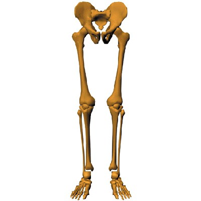
Consider the human skeleton:the skeleton of the lower limb and upper limb is represented by the belt and the free part. In the upper section, these are the bones: the chest girdle, the shoulder blades and the clavicle, the humerus and the bones of the forearm, the hand. Human bones of the lower limb include: the pelvic girdle (or paired pelvic bones), the thigh, the shin, and the foot. The bones of the free lower limb of a person, as well as the belts, are capable of supporting the weight of a person, therefore, they are so important for him. After all, in fact, only with the help of these connections can it be in a vertical position.
Pelvic girdle (paired pelvic bones)
The first component, which is the basis, forming the bones of the belt of the human lower limb, will be the pelvic bone.
Femur and Patella
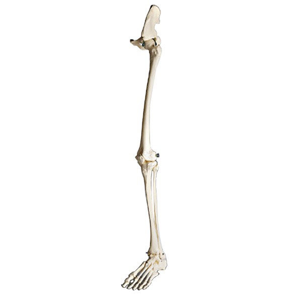
The bones of the belt of the lower limb of a person are notas mobile as the rest of it, which is called so - free lower limb. It consists of: thigh, shin and foot. The thigh, or femur, is a tubular bone. It is also the largest and longest of all the bones with which the human body is endowed. In its upper part, the femur connects to the pelvic girdle with the help of the head and a long thin neck. Where the cervix passes into the main part of the femur, there are two large bumps on it. It is here that the bulk of the muscles of the human lower limbs is attached. Down the femur becomes thicker. Here are two elevations, through which the thigh is connected, as a result, with the patella and the shin. The patella is a flat bone of a rounded shape, with which the knee is bent. The bones of the lower limb of a person, namely the thigh and patella, carry the following functions: the place of attachment of the bulk of the muscles located on the legs, and the possibility of bending the leg.
Shin
The human shin consists of two bones: tibial and fibula. They are located next to each other.

Human foot bones
Human foot bones are divided into three mainparts: bones of the tarsus, metatarsus and phalanges of the fingers. It is important to note that the foot is the free bones of a person's lower limb. The first ones include seven bones, the main ones being the bone, called the ram and forming the ankle, and the heel bone. The bones of the metatarsus are further located. There are only five of them, the first one is much thicker and shorter than the others. The toes of the foot are made up of bones called phalanges. The peculiarity of their structure is that the big toe contains 2 phalanges, the other fingers - three pieces each.
Anatomy of the joints of the human lower limbs. Sacroiliac joint, pubic symphysis
Just want to say that all the joints of the lower limbs are very large, compared with the joints of the upper limbs.
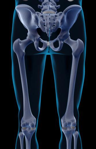
Human pelvis: large and small. Hip joint
It has already been described above that the bones of the belt belowhuman limbs are represented primarily by the pelvic bones. They, connecting with the sacrum and pubic symphysis, form a pelvis. It is, figuratively speaking, a ring that protects all organs, vessels and nerve endings inside from external influences. Distinguished large and small pelvis. In women, it is much wider and lower than in men. The fair sex is thought out to facilitate the generic process, so the pelvis has a more rounded shape and a large capacity.
Knee-joint
Imagine a human skeleton.The connection of bones in the form of joints is necessary for a person for the strength of the connection of bones and the creation of maximum mobility of all his limbs. An excellent example of such a connection is the knee joint. He, by the way, is considered the largest joint in the human body. And its structure is very complex: a knee joint is formed with the help of condyles, patella, tibial condyles. The entire joint is shrouded in reliable ligaments, which, along with ensuring the movement of the leg, keep it in the desired position. Thanks to him, not only standing, but also walking. The knee joint can produce various movements: circular, flexor and extensor.
Ankle joint
This joint serves to directly connect the foot and lower leg. Around there are numerous ligaments that provide a variety of movements and the necessary stability of the human body.
Plusphalangeal joints
The joints under study are interesting in their shape, according tocompared with other joints of the human lower limb. They look like a ball. Strengthening for them are bundles on the sides and on the sole of the foot. They can move, although the diversity of their movement does not differ: small leads to the sides, flexion and extension. Human foot consists of numerous (sedentary) joints and ligaments. With their help, and the movement is carried out, while the human body has the necessary support. So, we can conclude that the bones of the belt of the lower limb of a person are less mobile than the free bones of the same section. But the functions of this no less than those of none.
How do limbs develop with age?
We all know that during life certainThe human skeleton also undergoes transformations. The skeleton of the lower limb undergoes strong changes with age. The bones that develop on the basis of connective tissue have three stages of their change: connective tissue, cartilage and bone tissue.

Remember that the human skeleton is subject to differentinjuries in the form of injuries and even fractures. Since he is entrusted with the implementation of such a large number of important functions of the body, it must be protected. Eating right (food with sufficient calcium content helps strengthen the skeleton), lead an active lifestyle (physical education and sports), monitor your health (any disruption in the functioning of the skeleton to check with a competent specialist) - all this needs to be done by everyone. And then you will meet your old age vigorous, healthy and cheerful.