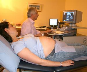
Fetal ultrasound is one of the methods of intrauterinemonitoring the state and development of the child. This procedure is based on the action of sound waves, whose oscillation frequency is not audible to the human ear. Like echoes, they are reflected from various tissues with varying strength, turning into a picture that is displayed on the monitor.
What does the fetal ultrasound:

- Sets the exact duration of pregnancy.
- Determines the number of embryos.
- Determines the place where the placenta was attached.
- Detects the presence of fibroids or other formations in the small pelvis, which, in one way or another, interfere with the favorable development of pregnancy.
- Time reveals the pathology of fetal development.
During pregnancy, the future mother will need to visit three planned ultrasound of the fetus.
The first is held for a period of 10 to 14 weeks.The main purpose of this procedure is to identify the presence of vestigial defects in the fetus (for example, hydrocephalus or Down Syndrome) and determine the date of birth. In case of detection of pathology, the doctor decides either to save the pregnancy, or to get rid of it (naturally, with the consent of the mother).
The second expert ultrasound of the fetus needs to be done ininterval between the 20th and 24th weeks. At this time, the fetus has already clearly formed all the organs, and this study is conducted to study their development. If any pathology is found, intrauterine treatment is used. Also, in the second study, the condition of the placenta and the amount of amniotic fluid are carefully analyzed.
The third ultrasound is performed for a period of 30 to 34 weeks. It also examines all internal organs of the fetus, assesses the condition of the placenta, uterus and amniotic fluid.

In addition to planned ultrasound, a doctor can also schedule an unscheduled study. The reason for its conduct can be:
- This study is performed to clarify the timing of pregnancy before: stimulation of labor, cesarean section, the conduct of abortion.
- The presence of some diseases of the mother (diabetes, arterial hypertension, preeclampsia, etc.), which can cause an intrauterine delay in the development of the fetus.
- Bleeding during pregnancy.
- If you suspect a multiple pregnancy.
- If there is a formation in the cavity of the small pelvis, which was detected by manual examination.
- To exclude an ectopic pregnancy.
- If you suspect a frozen pregnancy (fetal death).
- When suspected of low or polyhydramnios.
- To assess fetal anomalies identified earlier.
The whole procedure of ultrasound takes no more than 25 minutes.It is absolutely painless and safe for both the woman and the fetus. At the term of pregnancy less than 12 weeks, ultrasound is performed by a vaginal sensor, more than 12 - by a sensor, which is driven to the abdomen.
In recent years, future mothers have becomeuse a new type of diagnosis - 3d fetal ultrasound. This study is a three-dimensional ultrasound, which gives even more information about some of the defects of the fetus. In addition, 3D ultrasound allows the pregnant woman to see some parts of the baby's body and his face. Also, the entire procedure can be recorded on a digital medium.


