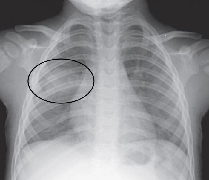
One of the radiation diagnostic methods for internalorgans is x-ray transmission, or radiography. The resulting image is applied to a hard disk, special film or paper.
Purpose of the survey
Radiography of the lungs is the most common and informative method of investigation. This diagnostic method allows to detect the presence of respiratory diseases:
- sarcoidosis;
- pneumonia (pneumonia);
- malignant neoplasms;
- tuberculosis;
- chest injuries;
- the presence of foreign objects;
- pneumothorax and other various pathological processes.
In order to prevent lung diseases in citizens,employed in hazardous industries (chemical industry, construction (bricklayers), mining (miners), etc.), once a year (more often if necessary), x-rays of the lungs are performed. What show research results in such cases?

The effect of radiation on the human body
Ray transmission is considered asradiation exposure, and some people refuse to perform this procedure. However, it is in vain, in medicine low-energy rays are used, the radiation dose is negligible, and the human body is exposed to them for a short period. A few years ago, scientists proved that even repeated radiographs (for medical reasons) are not capable of harming health. In some cases, this procedure is prescribed for pregnant women. Serious diseases that can be diagnosed with X-rays have more serious consequences than the minimum dose of radiation. As an alternative to conventional conventional X-rays, digital is available at present, with an even lower radiation dose.
Indications
Consider the symptoms in which the attending doctor prescribes an x-ray of the lungs. What will show a snapshot, the tactics of further patient management will depend on this.
- Periodic pain in the sternum.
- Dyspnea.
- High body temperature, which lasts a long time.
- Blood in the sputum.
- Long exhausting cough.
- A large amount of discharge of sputum.
- Dry cough.
For the purpose of prophylaxis, fluorography, or x-rays, is shown to all citizens at intervals of at least once every two years or more often in accordance with the recommendations of a medical professional.
Preparation and conduct of the procedure
The direction of the x-ray of the lungs is written outprepare for it? Preliminary preparation is not required. Before carrying out the procedure, it is necessary to remove the jewelry (chains, beads, necklaces) so that they do not distort the result. Immediately before the procedure, a medical professional will ask you to wear a special waist-skirt to protect the genitals from radiation. Next, the doctor chooses the desired projection (front, back, or sometimes the picture is taken in the supine position).

X-ray results
X-rayed lungs? What decryption shows, consider below:
- Diaphragm Defects.
- The presence of fluid in the pleural cavity. Exclude a tumor or pleurisy.
- A cavity in the lung indicates necrosis of the lung tissue. Diagnose tuberculosis, cancer or abscess.
- Small focal blackouts are signs of pneumonia, tuberculosis. Large - a tumor of the bronchi, metastases in the lungs.
- Small foci that occur very often are sarcoidosis or tuberculosis.
- A large round-shaped shadow - tuberculosis in the progression stage or a malignant neoplasm.
Except for the above, andother changes of the lung tissue and lungs that help make the correct diagnosis and prescribe treatment. Unfortunately, there are cases and a false result, or in cases of conducting research in the early stages of the disease, it can not be seen. In addition to X-rays, other diagnostic methods are used for accurate conclusions in addition to the results obtained, as well as the necessary laboratory tests are carried out.
X-Ray Dimming
X-ray showed spots on the lungs?The reasons for their occurrence may be: the wrong position of the patient during the procedure, poor-quality equipment, the presence of pathology. Exact decoding of radiography data can only be done by a doctor.
Formations in the form of white spots indicate the presence oftuberculosis, bronchitis, pneumonia, pathologies in the pleura, occupational diseases. If a person has had been ill with bronchitis, pneumonia, then spots can be found on the X-ray. They are regarded as residual manifestations of the disease, and they will disappear after a while.
If light spots are found in the upperparts of the lung, then diagnosed with tuberculosis, the main feature in the first stage of which is a light path, going from the place where there is an inflammatory process, to the root system. With timely and proper treatment, inflammation is reduced and the tissues undergo scarring. A dark spot appears in the picture instead of white.
If the snapshot x-ray shows that black spots are visible, this indicates aggravation andthe presence of chronic pneumonia. After a course of drug treatment and full recovery, the spots disappear. Dark formations can be the cause of malignant pathologies. Detection of dark spots in a practically healthy person indicates a long-term smoking, in children - a foreign body.
Does x-ray show pneumonia?
X-ray examination for pneumonia is both a method for detecting a disease and monitoring its course.

- global spotty formations on the entire surface of the lungs;
- Subtotal - completely all fields (exception - upper lobes);
- segmental - spots within a segment;
- small spotty formations up to 3 mm with limited margins.
В результате развития воспалительного процесса в Human lungs form fuzzy spots with blurred outlines and X-rays show inflammation of the lungs. The manifestation of spotted formations depends on the stage of the disease. More pronounced spots in advanced cases.
X-ray for bronchitis
Symptoms of the disease are similar to pneumonia.To confirm the diagnosis in case of a protracted course of the disease, certain types of examinations are prescribed, including X-rays, which will make it possible to assess the condition of the organs of the respiratory system and clarify the diagnosis.

- change in blood, according to laboratory tests;
- severe persistent dyspnea;
- prolonged increase in body temperature;
- suggestion of inflammation in the lungs;
- signs of obstruction.
According to the results of the study on x-rays, pay attention to the following points in the lungs:
- fuzzy contours;
- presence of root deformation;
- changes in the drawing;
- the presence of lamellar foci;
- areas of fluid accumulation.
Opinions of experts about the informativeness of X-rays in identifying the disease bronchitis were divided. However, this type of research is widely used in practical medicine.
X-ray in tuberculosis
If you suspect this serious disease, this type of examination of the lungs will allow you to exclude or confirm pathology.

- conduct various diagnostics of the disease;
- exclude other pathologies of the respiratory system, for example, pneumonia, cancer, abscess and others;
- determine the nature of damage to the lung tissue;
- see the prevalence of lesions;
- see the location of pathological foci.
Therefore, the question can x-ray show pulmonary tuberculosisanswer in the affirmative. However, this does not preclude additional manipulations to confirm the diagnosis accurately. X-rays reveal different types of tuberculosis:
- intrathoracic lymph nodes;
- disseminated;
- focal;
- infiltration;
- caseous pneumonia;
- fibro-cavernous;
- cirrhotic.
Does x-ray show lung cancer?
This disease is one of the terrible ailments.man in recent decades. Chest X-ray is considered a diagnostic method for detecting this pathology at the earliest stages of its development. Signs or symptoms of the disease can be:
- lethargy, constant sleepiness and weakness;
- performance at zero;
- regular fevers with apparent well-being;
- dyspnea;
- breathing with a whistle;
- a prolonged cough that does not respond to therapy;
- sputum with blood;
- lack of appetite;
- with bouts of cough, the presence of pain.
To exclude the disease, the doctor prescribes an examination. X-ray shows lung cancer necessarily, as this method is highly informative.

X-ray of the lungs in children
If your child was prescribed an x-ray, you should be familiar with the following points:
- Is there an alternative survey type?
- Is there a vital need for this procedure?
If in doubt, consult another specialist.
The younger generation in exceptional cases, prescribe X-rays. Basically, when this is the only manipulation, with the help of which it is possible to exclude or confirm the diagnosis.

Roentgenoscopy is an effective method of diagnosing various diseases and, in experienced hands, provides invaluable assistance to the medical community.


