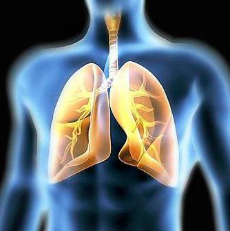
Определение границ легких имеет большое значение for the diagnosis of many pathological conditions. The ability to percussionally reveal the displacement of the thorax organs in one direction or another allows one to suspect the presence of a certain disease at the stage of examining the patient without using additional methods of investigation (in particular, radiographic ones).
How to measure the boundaries of the lungs?
Of course, you can use instrumentalmethods of diagnosis, to make an X-ray and evaluate how the lungs are located relative to the skeletal structure of the chest. However, it is best to do this without exposing the patient to radiation.

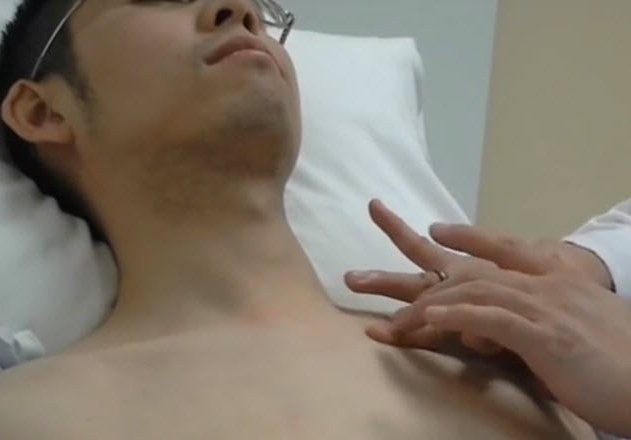
This study is being carried out as follows.One hand is placed palm on the study area, two or one finger of the second hand strikes the middle finger of the first (plessimetre), like a hammer on the anvil. As a result, you can hear one of the options percussion sound, which have already been mentioned above.
Percussion is comparative (sound is evaluated in symmetrical areas of the chest) and topographic. The latter is just designed to determine the boundaries of the lungs.
How correctly to conduct topographic percussion?
The finger-plessimeter is set to the point, with(for example, when determining the upper border of the lung over the anterior surface, it starts over the middle part of the clavicle), and then shifts to the point where approximately this measurement should end. The border is defined in the area where the pulmonary percussion sound becomes blunt.

Upper bound
The position of the apex of the lung is assessed as from the front,so and behind. On the anterior surface of the chest, the clavicle serves as a guide, on the back - the seventh cervical vertebra (it has a long spinous process along which it can easily be distinguished from other vertebrae).
The upper limits of the lungs are normal as follows:
- In front of the level of the clavicle 30-40 mm.
- The back is usually at the same level as the seventh cervical vertebra.
The study should be performed as follows:
- At the front finger-plessimet is placed above the clavicle (approximately in the projection of its middle), and then shifts upwards and to the inner part, until the percussion sound becomes blunt.
- Behind the study begin from the middle of the awningscapula, and then the finger-plessimeter is shifted upwards so as to be on the side of the seventh cervical vertebra. Percussion is performed until a dull sound appears.

Displacement of the upper border of the lungs
The displacement of the boundaries upwards occurs because of the excessairiness of lung tissue. This condition is typical for emphysema - a disease in which the walls of the alveoli become overextended, and in some cases, their destruction with the formation of cavities (bulls). Changes in the lungs with emphysema are irreversible, the alveoli swell, the ability to subside is lost, the elasticity is sharply reduced.
The boundaries of the human lungs (in this case the boundariestops) can be shifted and down. This is due to a decrease in airiness of the lung tissue, a condition that is a sign of inflammation or its effects (proliferation of connective tissue and wrinkling of the lung). Limits of the lungs (upper), located below the normal level, is a diagnostic sign of pathologies such as tuberculosis, pneumonia, pneumosclerosis.
Bottom line
To measure it you need to know the basictopographic lines of the chest. The method is based on moving the researcher's hands along these lines from top to bottom until the pulmonary percussion sound changes to a blunt sound. It should also be aware that the front left lung border is not symmetrical to the right one due to the presence of a pocket for the heart.

On the side, threeaxillary lines - anterior, middle and posterior, which begin from the anterior margin, the center and the posterior edge of the armpit, respectively. Behind, the edge of the lungs is defined relative to the line descending from the angle of the scapula, and the line located to the side of the spine.
Displacement of the lower border of the lungs
Следует отметить, что в процессе дыхания объем this body is changing. Therefore, the lower border of the lungs are normally displaced 20-40 mm up and down. A stable change in the position of the border indicates a pathological process in the chest or abdominal cavity.

Вверх границы легких перемещаются обычно due to the wrinkling of the latter (pneumosclerosis), the fall in the share as a result of bronchial obstruction, accumulation of exudate in the pleural cavity (as a result of which the lung collapses and contracts to the root). Pathological conditions in the abdominal cavity are also capable of shifting the pulmonary boundaries to the top: for example, fluid accumulation (ascites) or air (with perforation of the hollow organ).
Limits of lungs are normal: table
Lower boundaries in an adult | ||
Field of study | Right lung | Left lung |
Line at the lateral surface of the sternum | 5 intercostal spaces | - |
Line descending from the middle of the clavicle | 6th rib | - |
The line originating from the anterior margin of the axilla | 7th rib | 7th rib |
The line from the center of the axilla | 8th edge | 8th edge |
Line from the back edge of the axilla | 9th rib | 9th rib |
Line descending from the angle of the blade | 10th rib | 10th rib |
Line to the side of the spine | 11 thoracic vertebra | 11 thoracic vertebra |
The location of the upper pulmonary boundaries is described above.
Change in the index depending on the build
In asthenic lungs are elongated in the longitudinaldirection, therefore, they often fall slightly below the generally accepted norm, ending not in the ribs, but in the intercostal spaces. For hypersthenics, on the contrary, a higher position of the lower boundary is characteristic. Their lungs are wide and flattened in shape.
How are the pulmonary borders of the child?
Strictly speaking, the boundaries of the lungs in children are practicallycorrespond to those of an adult. The tops of this organ in children who have not yet reached the preschool age are not defined. Later, they are detected anteriorly 20–40 mm above the middle of the clavicle, and behind, at the level of the seventh cervical vertebra.

The location of the lower bounds is discussed in the table below.
The boundaries of the lungs (table) | ||
Field of study | Age up to 10 years | Age over 10 years |
Line running from the middle of the clavicle | Right: 6 edge | Right: 6 edge |
Line originating from the center of the armpit | Right: 7-8 edge Left: 9th rib | Right: 8 edge Left: 8 rib |
Line descending from the angle of the blade | Right: 9-10 rib Left: 10 edge | Right: 10 edge Left: 10 edge |
The reasons for the shift of pulmonary boundaries in children up or down relative to normal values are the same as in adults.
How to determine the mobility of the lower edge of the body?
Выше уже говорилось о том, что при дыхании нижние boundaries are shifted relative to normal values due to the expansion of the lungs during inhalation and a decrease in exhalation. Normally, such a shift is possible within 20-40 mm upwards from the lower border and as much downwards.
Определение подвижности осуществляют по трем the main lines, starting from the middle of the clavicle, the center of the axilla and the angle of the scapula. The study is conducted as follows. First, determine the position of the lower boundary and make a mark on the skin (you can handle). Then the patient is asked to take a deep breath and hold the breath, then again find the lower limit and make a mark. And in conclusion, determine the position of the lung during maximum exhalation. Now, focusing on the marks, one can judge how the lung shifts relative to its lower boundary.
In some diseases, the mobility of the lungsnoticeably reduced. For example, this occurs during adhesions or a large amount of exudate in the pleural cavities, loss of elasticity by the lungs during emphysema, etc.
Difficulties during topographic percussion
Данный метод исследования непрост и требует certain skills, and even better experience. Difficulties arising from its use, usually associated with improper execution technique. As for the anatomical features that can create problems for the researcher, this is mainly pronounced obesity. In general, it is easiest to perform percussion on asteniki. The sound is clear and loud.

- Know exactly where, how and exactly what boundaries you need to look for. Good theoretical training is the key to success.
- Move from clear to blunt sound.
- The finger-gauge should lie parallel to the defined boundary, but should move perpendicular to it.
- Hands should be relaxed. Percussion does not require significant effort.
And, of course, experience is very important. Practice gives confidence in their abilities.
Summarize
Percussion - very important in the diagnostic planresearch method. It allows to suspect many pathological conditions of the chest organs. The deviation of the boundaries of the lungs from normal indicators, impaired mobility of the lower edge - the symptoms of some serious diseases, timely diagnosis of which is important for the full treatment.


